Biological Imaging & Functional Morphology
Our imaging research deals with initially visualizing newly discovered species, quantifying morphological variation, and reconstructing lost phenotypes. While some of these projects involve traditional methods such as light microscopy, alternative imaging techniques offer highly informative new avenues for data collection. Through X-ray computed tomography (CT-scanning), it is possible to create three-dimensional reconstructions of specimens from several hundred individual 2D X-ray images. The resulting reconstructions provide improved visibility of external structures and internal tissue (including in some amber fossil specimens). Certain specimens also lend themselves well to laser confocal microscopy, a process that yields high depth detail, something that is often prohibitive in light microscope scenarios. We are especially interested in blending all available imaging resources to extract the maximum amount of information from long-extinct organisms.

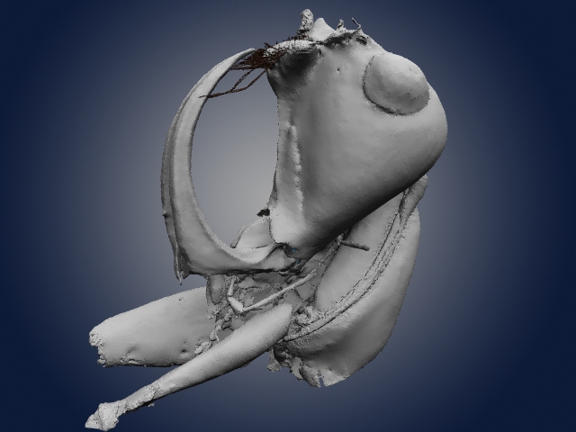
CT-scan derived 3D reconstruction of Leptomyrmex neotropicus from 20 million year old Dominican amber.

Ventral view of Leptomyrmex neotropicus in Dominican amber – structures resolved in orange are musculature.

Alternate views of early fossil evidence for social behavior – CT reconstruction of eleven ant workers trapped in 99 million year old Burmese amber.

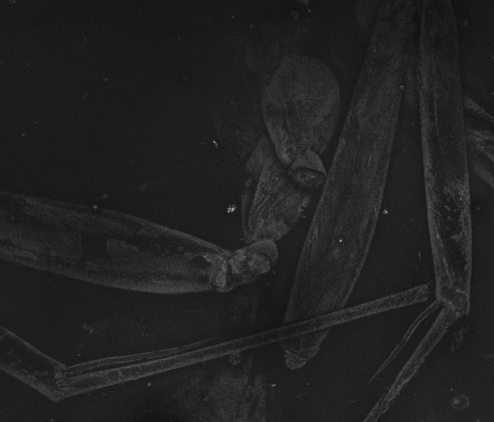
Lateral photomicrograph of Zigrasimecia tonsora entombed in 99 million year old Burmese amber.
3D rendering of the head, mandibles, and prothorax of the extinct and Haidomyrmex scimitarus.
Laser confocal image of the mid and hind legs of a 52 million year old dolichoderine ant preserved within Cambay amber from Gujarat, India.

A 2D X-ray image of the mandibles and head capsule of Zigrasimecia tonsora from 99 million year old Burmese amber.
Much of this work is done at the American Museum of Natural History Microscopy and Imaging Facility, however we have also utilized a powerful imaging beam line at the Argonne National Laboratory Advanced Photon Source and microCT instruments at the Cornell University Biotechnology Imaging Center.